Musculoskeletal ultrasound is the painless alternative to diagnose ailments.
Dr. Keith Feder, Dr. Carol Frey and the West Coast Center for Orthopedic Surgery and Sports Medicine routinely uses musculoskeletal ultrasound to diagnose and manage a number of ailments and conditions for our patients in Manhattan Beach, Los Angeles and surrounding areas. Using this technique often eliminates the need for an MRI or CT scan.
Musculoskeletal (MSK) ultrasound is a powerful and painless tool used to provide real-time images of muscles, tendons, ligaments, nerves, and cartilage throughout the body.
MSK ultrasound is particularly helpful in the diagnosis of orthopedic and sports injuries, such as rotator cuff tears, and chronic conditions, such as rheumatoid arthritis. Sometimes, pain or injury is triggered by movement, which cannot be captured in a static image. Ultrasound is performed in real time and can provide unique information that cannot be detected by any other imaging method. It has the added advantage of being radiation-free.
MSK ultrasound is used to diagnose a wide range of injuries and chronic conditions, including muscle tears, tendonitis, bursitis, joint problems, rheumatoid arthritis, and masses such as tumors or cysts.
MSK ultrasound is also used by musculoskeletal radiologists to guide injections and pain management procedures, because it allows for real-time visualization of the needle as well as the joints and soft tissue.
How do I prepare for the test?
On the day of the test you should wear comfortable clothes, and you may be asked to change into a gown when you arrive at the ultrasound exam site. No special preparation is needed.
What will happen during the test?
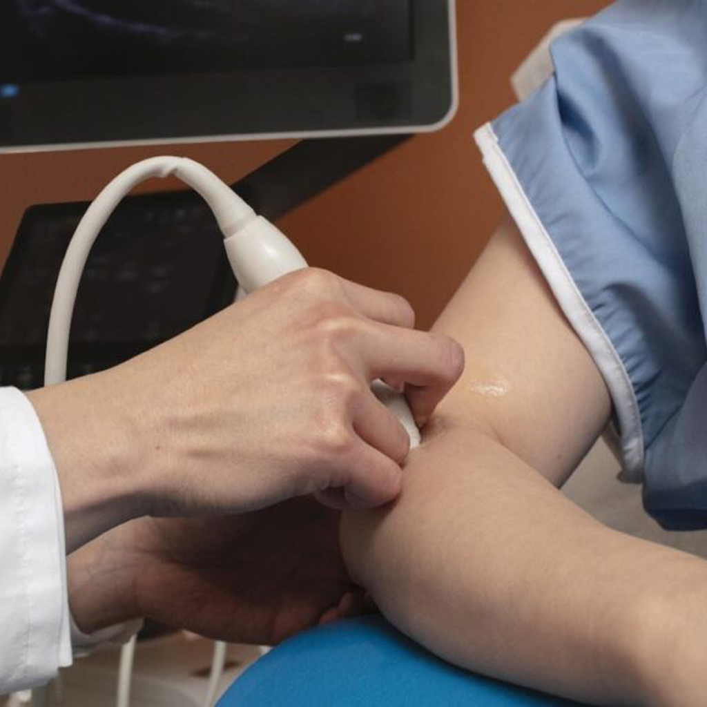
During a musculoskeletal ultrasound the sonographer may ask you to sit on the exam table, on a swivel chair, or lie face up or face down. He/she will cover the skin over the area to be examined with a small amount of warm gel to prevent air pockets from forming between the transducer and skin. The sonographer will then glide the transducer over your skin to capture the images of the tissues below, which the transducer then sends to a computer.
During the ultrasound, we may ask you to move the joint or limb being examined in order to evaluate the function of the joint, muscle, ligament, or tendon.
An ultrasound exam is typically painless, and takes about 20-30 minutes to complete.
Are there any risks?
Ultrasound does not require the use of ionizing radiation, special dyes, or anesthesia and is a safe diagnostic tool with no known risks or side effects.
After the test
After the exam you can immediately resume your normal activities. A radiologist will analyze the ultrasound images and will share the results with the doctor who requested the exam. Your doctor will then discuss the results with you.

Some of the Benefits of Musculoskeletal Ultrasound
There are several benefits to using musculoskeletal ultrasounds to diagnose and manage your condition.
- We are able to study your bone structures and soft tissues so that we can make accurate evaluations.
- Ultrasound is comfortable for you and many patients appreciate avoiding the uncomfortable and claustrophobic nature of MRIs and scans.
- The images we get from the Ultrasound are live and in real time. This helps us move you quicker to the right treatments.
- When treating you with PRP therapy, bone marrow stem cell therapy or other injections, this helps us ensure the accuracy of our needle placement.
- Musculoskeletal Ultrasounds are usually less expensive than CT scans or MRIs.

