Focus on Foot and Ankle
Carol Frey, MDa,*, Alessandra Falk, MDb
KEYWORDS
- Metabolic bone health
- Female
- Stress fracture
- Female triad
- RED-S
- Lactation
- Bone marrow density
KEY POINTS
- Low energy availability—the female triad or relative energy deficiency in sport—drives premature bone loss during adolescence when most bone mass is acquired.
- Menstrual dysfunction occurs in up to 66% of female athletes versus 2% to 5% of the general population, and untreated amenorrhea can decrease bone mass.
- Combined oral contraceptives may normalize bleeding without restoring bone health; restoring caloric intake, reducing overtraining, and—if needed—transdermal 17β-estradiol are preferred.
- Stress fracture are often the first sign of compromised bone health and should prompt DXA scan interpreted by Z-score, not T-score, in adolescents and premenopausal women.
- Lactating patients demineralize trabecular bone to meet milk calcium demands, making the lactating athlete an under-recognized fracture risk group.
Abbreviations
- BMD – bone mineral density
- BMI – body mass index
- COCs – combined oral contraceptives
- DXA – dual-energy X-ray absorptiometry
- FAST – Female Athlete Screening Tool
- LEA – low energy availability
- LEAF-Q – Low Energy Availability in Females Questionnaire
- REDS – relative energy deficiency in sport
INTRODUCTION
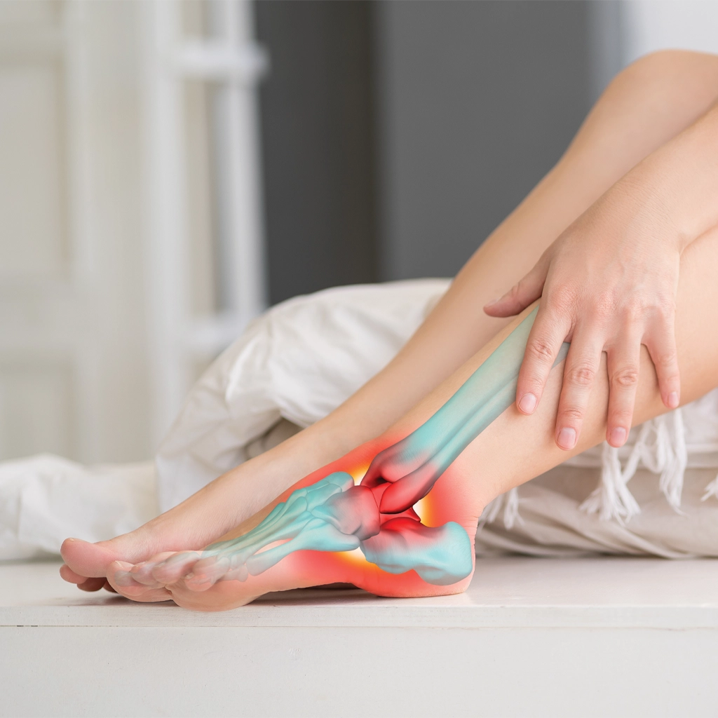
Metabolic [GS1.1][Cf1.2]bone health is required for the overall well-being of the female patient. Female athletes and young females are of particular interest, as their skeletal systems are still developing and are sensitive to the hormonal and physical stress of training and diet. Hormonal status, mechanical loading, and nutrient intake interact in a significant way on bone structure, especially in major weight-bearing areas of the foot and ankle. An early sign of deficient bone health can be a stress fracture in the lower extremity, particularly in the metatarsals, navicular, calcaneus, and distal tibia. Stress fractures occur with higher frequency in females [ND2.1] with risk factors such as low bone mineral density (BMD). When low bone mineral density occurs along with menstrual irregularities and relative energy deficiency, it completes the Female Triad or REDS (relative energy deficiency in sport).1–3Any discussion of female BMD and metabolic bone health must include the Female Triad. The Female Triad may lead to decreased bone marrow density or bone thickness. In a young female, she may ultimately not acquire enough bone during critical periods. This can lead to osteoporosis, poor metabolic bone health and skeletal degradation as an adult.
The Triad and Bone Health
Three components are central to the concept of the Female Triad: eating disorders, irregular menstrual cycle, and a decrease in bone density. More recently, the Female Triad has been considered too restrictive in its description of a disorder that starts with low energy availability (LEA). One of the criticisms has been that it always included eating disorders. In 2005, the IOC put out a consensus statement4,5that described RED-S. RED-S is a broader description used to describe a physiologic disorder in athletes. RED-S syndrome includes menstrual dysfunction, bone health, relative energy deficiency, and metabolic health. The syndrome does not need to include all components for the athlete, usually female, to be diagnosed with the disorder. The athlete will present with an energy deficiency resulting from an imbalance between energy intake and the energy required for sports performance. The Female Triad can occur in any female who is of menstrual age. RED-S is related to energy balance and a lack of sufficient energy to maintain bone health, menstrual function, and other physiologic functions that can impact sports performance. It is not a triad, but rather a spectrum. From the viewpoint of an orthopedic surgeon, any of the conditions that make up the Triad and RED-S can lead to premature bone loss and a disruption of metabolic bone health. In females, most bone mass formation is in the late teen years. Young women with the Female Triad or RED-S may ultimately fail to acquire sufficient bone during this critical time, leading to osteoporosis and poor skeletal health in adulthood.
Relative Energy Deficiency in Sport and Bone Health
RED-S significantly impairs bone metabolic health in young female athletes by disrupt
ing the hormonal and physiologic processes necessary for normal bone remodeling and repair. RED-S can lower levels of Insulin-like Growth Factor-1 and Leptin, both necessary anabolic mediators of bone growth. The energy deficiency suppresses the hypothalamic-pituitary-ovarian axis, resulting in decreased estrogen production, particularly estradiol, which is essential for inhibiting bone resorption and promoting bone formation. Consequently, bone turnover becomes unbalanced—favoring resorption over formation—leading to decreased BMD and increased risk of stress fractures, especially in trabecular-rich bone such as the spine, pelvis, and calcaneus. In adolescents and young women, RED-S can suppress the acquisition of peak bone mass.4–6
RED-S also affects other metabolic pathways involved in bone health, which can impair bone microarchitecture and lead to higher rates of stress injuries compared to eumenorrheic athletes with good energy availability.2,3,7,8 Even in the absence of amenorrhea, any menstrual dysfunction (e.g., luteal phase defects) can compromise bone health. RED-S is a serious condition that should be screened for during an annual or preseason physical by the team physician who is often an orthopedic surgeon.4,5,9
Menstrual Function and Bone Health
Menstrual function is an important component of the Female Triad and RED-S. Some studies of female high school athletes have reported menstrual dysfunction present in more than 50%.10–14 Athletes with menstrual irregularities were noted to be 3 times more likely to sustain an injury and have a greater reported time off from play when injured than those without irregularities. Problems with menstruation include no menstruation (amenorrhea) and irregular menstruation. Amenorrhea can be primary (failure of menstruation to begin) and secondary amenorrhea (cessation of the cycle in a female who previously had a regular cycle). Secondary amenorrhea is defined as more than three to 6 months passing without a menstrual cycle. Primary amenorrhea is a delayed onset of menstruation.11–16 No periods or irregular periods have been reported to occur in up to 66% of female athletes,1–3,9,10,17–22 as compared. to 2% to 5% of women in the general population.11–14 If amenorrhea is left untreated, the female will continue to lose bone mass, which can be up to 3% per year. Lack of menstruation for even 6 months during adolescence can result in permanent bone loss.11–14
Decreased Bone Mineral Density
Decreased BMD has been reported in amenorrheic athletes at rates as high as 22% to 50% compared to 12% in the general population.23 Low body weight, particularly a body mass index (BMI) lesser than 18.5 kg/m2, is strongly associated with hypoestrogenism, decreased mechanical load on the skeleton, and nutritional deficits. These all combine to contribute to reduced bone formation and increased bone resorption. Female athletes with low BMI or excessive leanness, even without an eating disorder, are at heightened risk for LEA. The effects on the hypothalamic-pituitary-ovarian axis can result in menstrual dysfunction and suppressed estradiol levels, which compromise bone health.4–6,24 There are only a limited number of studies that look at the bone density of young female athletes. Most studies look at older women who are menopausal. In a review of 437 exercising women, Gibbs found that a cumulative number of Triad risk factors were associated with an increased risk of low BMD. Gibbs defined low BMD as a dual-energy X-ray absorptiometry (DXA) Z score of smaller than or equal to minus 2.23 A 2006 survey by Nichols10 examined 170 high school female athletes, and approximately 22% had low bone marrow density.
Oral Contraceptives and Bone Health
The use of birth control pills, particularly combined oral contraceptives (COCs), can influence BMD and bone health in young females. Estrogen plays a crucial role in bone accrual during adolescence and early adulthood, when peak bone mass is achieved. By suppressing endogenous estrogen production through negative feedback on the hypothalamic-pituitary-ovarian axis, COCs may result in lower overall estrogen exposure compared to natural menstrual cycles. This effect may be exacerbated in females using low-dose COCs (containing <30 μg of ethinyl estradiol), resulting in reduced bone mass accrual in young females. Because females typically reach peak bone mass in their early twenties, decreased bone acquisition during this period may increase the lifetime risk of osteoporosis and stress fractures. In young athletes or those with low energy availability (The Triad or RED-S), COCs do not adequately replace the bone-protective effects of natural estrogen. While they may induce withdrawal bleeding, they do little to correct underlying metabolic and hormonal factors necessary for optimal bone metabolic health.
Miller and colleagues25 reported that COC-treated amenorrheic athletes had low spinal BMD that persisted, in contrast to patients who resumed normal menstrual function through weight gain and proper diet alone. The best way to restore normal menstrual function in young females is to restore adequate caloric intake and address overtraining. If hormonal therapy is required, transdermal estradiol may be recommended. As mentioned, estrogen plays a critical role in maintaining and accumulating bone mass. However, the synthetic ethinyl estradiol found in most COCs, especially at low doses of 20 to 30 μg, is not equivalent in its physiologic effects to endogenous 17β-estradiol. The practice of prescribing COCs to regulate menses in athletes may give the appearance of hormonal balance but fails to restore the hypothalamic- pituitary-ovarian axis or improve bone metabolic health.7,8,25 In a randomized controlled trial, Ackerman and colleagues7,8 found that transdermal 17β-estradiol significantly improved bone mass accrual in oligo-amenorrheic athletes with a menstrual cycle that is infrequent or absent.
In contrast, COCs did not offer such a benefit. COCs also suppress hepatic production of insulin-like growth factor 1, an essential anabolic hormone for bone formation. Of note, for mature menstruating women, COCs may have a neutral or somewhat beneficial effect on BMD.26–36
Lactation and Bone Health
While the athlete has a complex relationship with metabolic bone health that must be managed appropriately, there is another subset of the female population that may be at risk for low bone mass and fracture risk, namely the lactating patient.
During lactation, there is a state of low estrogen that is coupled with a loss of calcium from bones. Daily loss of calcium in breastmilk has been estimated to range from 280 ±400 mg, although daily losses as significant as 1000 mg calcium have been reported. This can vary across women, ethnic groups, and duration of lactation. The female body compensates for these calcium requirements by demineralization of the skeleton.7,8 This resorption is mainly seen in the trabecular bone due to the increased surface area that osteoclasts can readily absorb.
The decrease in bone mineral density is typically clinically silent in the average lactating patient. Few studies in the literature discuss the incidence of fractures during the lactation period. Kovacs and colleagues identify a host of factors that have been noted to contribute to lactation-related fractures, including hormonal, nutritional, pharmacologic, gastrointestinal, renal, connective tissue disorders, rheumatologictions, or genetic disorders.8 A relevant factor in the exhaustive list the authors provide is mechanical factors, including excess exercise. When combining the increased bone resorption seen in the lactating female with the previously described bone changes that occur in the female athlete, it stands to reason that a lactating female athlete is at higher risk for a fracture.
Given the significant energy commitment that is demanded by the lactating female, due to intrinsic factors of producing milk and the extrinsic factors of taking care of an infant, the incidence of high-performance athletes who are lactating is unsurprisingly not captured by the literature. One could imagine, however, that even seemingly modest exercise would stress an otherwise weakened bone, leading to stress fractures or a stress reaction of the bone. However, could the time demands of infant care may prevent repetitive exercise or deter these patients from seeking care until the pain is intolerable, thereby limiting their presence in the clinician’s office and studies of fractures.
Taken to its extreme, pregnancy and lactation-associated osteoporosis (PLO) is described in the endocrinology literature as having an incidence of 460 cases per million childbirths in Japan and 6.8 per 100,000 pregnancies in the United Kingdom. These numbers are increased from previous estimates, indicating an increase in awareness of this condition. PLO is not well understood but is assumed to be based upon the decrease in bone density that is combined with other factors, including history of activity, family history, and genetic factors. Vertebral fractures are the most commonly reported fractures, but other fracture sites have been reported as well.37 Typically, these fractures are treated nonoperatively. Current efforts are being made to increase awareness of this condition to guide patients most appropriately.
Studies suggest that the decreased bone mineral density recovers gradually at the conclusion of lactation. The extent to which this occurs is not clear and is debated in the literature. An extensive cohort observational study in the United States concluded that a history of lactation was not related to fracture risk or decreased bone density over time.38 By contrast, a study out of South Korea demonstrated that prolonged breastfeeding for more than 1 year per child demonstrated a deleterious effect on lumbar bone density.26 To further complicate the issue, some studies even indicate a protective effect of breastfeeding against osteoporosis.17 The extent to which this applies to foot and ankle pathology has yet to be determined and could be the subject of further studies.
Stress Fractures Can be the First Sign of Compromised Bone Health
The young female with symptoms of the Female Triad or RED-S often presents with a stress fracture of the foot and ankle. An important part of the workup for these patients involves evaluating bone density, especially if the patient has had more than 2 stress fractures or evidence of LEA.7,8 The DXA or DEXA is the gold standard for assessing BMD. While DXA is commonly used for osteoporosis screening in postmenopausal women, its use in adolescents and premenopausal women, particularly athletes or those with menstrual irregularities, also plays an important role in identifying low bone mass and fracture risk.
When evaluating the results of the DXA, keep in mind that the normal values for the DXA scan were established using Caucasian, postmenopausal women. Therefore, the same values should not be used when assessing adolescents and premenopausal females. Unlike older adults, where the T-score is used, DXA interpretation in young and premenopausal women relies on the Z-score, which compares a patient’s BMD to age-matched, sex-matched, and ethnicity-matched data. A Z-score of 2.0 or lower is considered below the expected range for age and suggests bone loss from secondary causes, such as hormonal dysfunction, nutritional deficiency, or chronic energy imbalance. All these secondary causes are common in young females with the Female Triad or RED-S.1,6–8,11–14,24
In young menstruating females, especially those at risk for RED-S or the Female Triad, DXA should be considered when there is a history of stress fractures, amenorrhea lasting more than 6 months, or signs of LEA. The scan typically includes measurements at the lumbar spine and femoral neck. Some DXA protocols also include measurements of the total body or distal radius. Additionally, serial DXA scans can help monitor changes in BMD over time, particularly during treatment or recovery periods from conditions such as the Female Triad, RED-S, or other causes of decreased BMD.
The International Society for Clinical Densitometry advises against using the term osteopenia or osteoporosis in premenopausal women and adolescents based on BMD alone. Instead, a Z-score lss than 2.0, accompanied by a clinically significant fracture history (such as vertebral fractures, 2 or more long-bone fractures by age 10, or 3 or more long-bone fractures by age 19), may justify a diagnosis of low bone mass for age.7,8
Treatment/Clinical Care Points
Early recognition and treatment of RED-S or the Female Triad involves multidisciplinary management, which is critical to prevent long-term consequences to bone health. The practitioner must ask questions about the menstrual cycle, weight, training outside of practice, use of BCP, and diet. The use of questionnaires or screening tools is often employed.39
The most widely validated screening tool for identifying (LEA) is the Low Energy Availability in Females Questionnaire (LEAF-Q). Melin40 developed the LEAF-Q and designed it for physically active women aged 15 to 35. The LEAF-Q evaluates risk in 3 main areas: menstrual function, gastrointestinal symptoms, and injury history, with a particular focus on stress fractures. A total score of 8 or more indicates a high risk of LEA and related conditions such as RED-S. The questionnaire has shown a sensitivity of 78% and a specificity of 90%.40 Another valid questionnaire is the Female Athlete Screening Tool (FAST), developed to assess disordered eating behaviors and attitudes among adolescent and collegiate female athletes. The FAST is a 33-item questionnaire.
Increasing caloric intake and body weight can help stimulate the body to normalize menstrual cycles. Typically, athletes need to consume a minimum of 2000 kcal/day, although their energy needs may far exceed that amount. Inadequate caloric consumption disrupts metabolic homeostasis and impairs bone remodeling. Supplementation of specific nutrients is recommended to support bone metabolic health. The recommended dose of calcium is 1300 mg/day for adolescents, and vitamin D is 600 to 1000 IU/day. If the amounts of these supplements are insufficient, it will impair mineralization and increase the risk of stress fractures. Protein intake supports bone matrix formation and is often underconsumed in restrictive eating patterns. Barrack reported that adolescent female runners with lower intakes of energy, calcium, and vitamin D had significantly reduced BMD at key skeletal sites, including the lumbar spine and pelvis.38
Resistance training has a well-documented positive impact on bone metabolic health in females, particularly during adolescence and early adulthood when peak bone mass is established. Mechanical loading from resistance exercise stimulates osteogenesis by applying strain to bone tissue, thereby enhancing BMD and improving bone geometry and strength. Unlike aerobic activity alone, resistance training produces localized stress on bone-tendon attachment sites, promoting remodeling and growth of cortical and trabecular bone.20,22,41
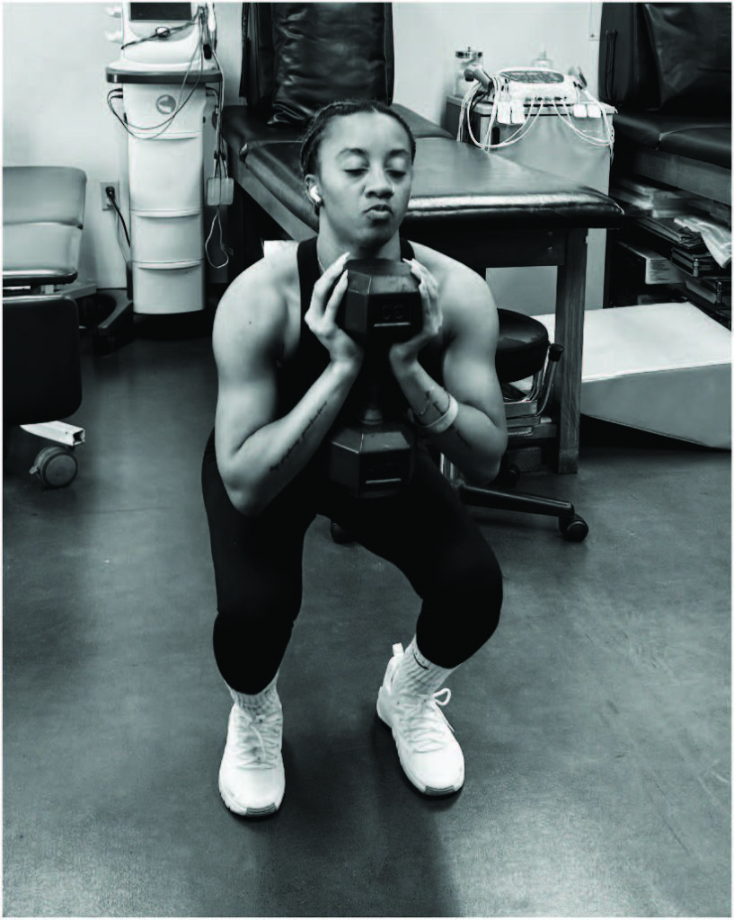
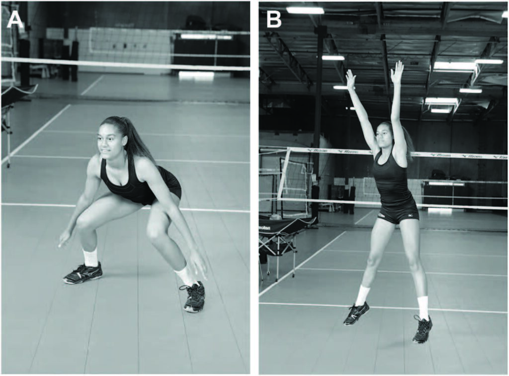
Research has shown that moderate-to high-intensity resistance training (approximately 70%–85% of one-repetition maximum) performed at least twice a week is effective in increasing or maintaining bone density in premenopausal women. Recommended exercises target major weight-bearing and postural muscle groups, including the lower body (eg, squats, lunges, and deadlifts), upper body (eg, shoulder press and push-ups), and core (eg., planks and medicine ball throws) (Fig. 1). Full-body dynamic exercises, such as step-ups and jump squats, provide the added benefit of impact loading, which further stimulates bone formation via neuromuscular pathways (Fig. 2A and B).41
Resistance training is a safe and critical for maintaining skeletal health in females. Unfortunately, strength and conditioning programs are often underemphasized in training protocols for female athletes, especially in high school. Its benefits are most significant when combined with adequate energy intake, calcium, and vitamin D, as well as menstrual regularity. Resistance training is a powerful tool for females to protect against early bone loss and reduce their risk of fractures later in life.
CLINICS CARE POINTS
- Early recognition of RED-S is critical to preserve bone health.
- To recognize RED-S, medical personel and trainers should feel comfortable asking females about menstrual function, diet and injury history–especially stress fractures.
- BMI < 18.5 is strongly associated with low estrogen, decreased load on the skeleton and poor bone health.
- A stress fracture of the lower extremity is often an early sign of poor bone health and low bone mineral density.
- Moderate to high instenisty resistance training is effective in increasing/maintaining bone density in females.
DISCLOSURE
C. Frey. Ossur, Medical Advisory. Pacira Pharmaceuticals Inc., Medical Advisory.
REFERENCES
- Nattiv A, Loucks AB, Manore MM, et al. American College of Sports Medicine position stand. the female athlete triad. Med Sci Sports Exerc 2007;39(10):1867–82.
- Tenforde AS, Barrack MT. Osteoporosis in the female athlete: pathophysiology, clinical presentation, and treatment options. Clin Sports Med 2016;35(3):367–84.
- Tenforde AS, Frederickson M. Influence of sports participation on bone health in the young athlete: a review. Pharm Manag PM R 2011;3(9):861–7.
- Mountjoy M, Sundgot-Borgen J, Burke L, et al. IOC consensus statement on RED- S: 2018 update. Br J Sports Med 2018;52(11):687–97.
- Mountjoy M, Sundgot-Borgen J, Burke L, et al. The IOC consensus statement: beyond the female athlete triad—relative energy deficiency in sport (RED-S). Br J Sports Med 2014;48(7):491–7.
- Misra M, et al. Evaluation and management of low bone density in children and adolescents. Pediatrics 2016;138(6):e20162300.
- Ackerman KE, Nazem T, Chapko D, et al. Bone microarchitecture is impaired in adolescent amenorrheic athletes compared with eumenorrheic athletes and nonathletic controls. J Clin Endocrinol Metab 2011;96(10):3123–33.
- Ackerman KE, et al. Transdermal 17β-Estradiol improves bone outcomes in oligo- amenorrheic athletes: a randomized controlled trial. J Clin Endocrinol Metab 2019;104(9):3590–600.
- De Souza MJ, et al. 2014 Female athlete triad coalition consensus statement on treatment and return to play. Br J Sports Med 2014;48(4):289–92.
- Nichols JF, et al. Prevalence of the female athlete triad syndrome among high school athletes. Arch Pediatr Adolesc Med 2006;160(2):137–42.
- Thein-Nissenbaum J, Carr KE. Female athlete triad syndrome in the high school athlete. Phys Ther Sport 2011;12(3):108–16.
- Thein-Nissenbaum J, Rauh MJ, Carr KE, et al. Associations between disordered eating, menstrual dysfunction, and musculoskeletal injury among high school athletes. J Orthop Sports Phys Ther 2011;41(2):60–9.
- Thein-Nissenbaum J, Rauh MJ, Carr K, et al. Menstrual irregularity and musculoskeletal injury in female high school athletes. J Athl Train 2012;47(1):74–82.
- Thein-Nissenbaum J. Long-term consequences of the female athlete triad. Maturitas 2013;75(2):107–12.
- Warren MP, Chua AT. Exercise-induced amenorrhea and bone health in the adolescent athlete. Ann N Y Acad Sci 2008;1135(1):244–52.
- Warren MP, Goodman LR. Exercise-induced amenorrhea and bone health in the adolescent girl. Ann N Y Acad Sci 2003;997:251–7.
- De Souza MJ, Ricker EA, Mallinson RJ, et al. Bone mineral density in response to increased energy intake in exercising women with oligomenorrhea/amenorrhea: the REFUEL randomized controlled trial. Am J Clin Nutr 2022;115(6):1457–72.
- Drinkwater BL, Nilson K, Chesnut CH III, et al. Bone mineral content in amenorrheic and eumenorrheic athletes. N Engl J Med 1984;311(5):277–81.
- Elliott-Sale KJ, et al. Methodological considerations for studies of menstrual cycle and oral contraceptive effects on exercise performance: insights from the TEAM study. Sports Med 2020;50(4):671–83.
- Kohrt WM, Bloomfield SA, Little KD, et al. Physical activity and bone health. Med Sci Sports Exerc 2004;36(11):1985–96.
- Nazem TG, Ackerman KE. The female athlete triad. Sports Health 2012;4(4): 302–11.
- Specker B, Binkley T. Randomized trial of physical activity and calcium supplementation on bone mineral content in 3- to 5-Year-Old children. J Bone Miner Res 2005;20(4):553–60.
- Gibbs JC, Williams NI, De Souza MJ. Definition and discussion of the etiology of the female athlete triad. Med Sci Sports Exerc 2014;46(1).
- Misra M, Klibanski A. Endocrine consequences of anorexia nervosa. Lancet Diabetes Endocrinol 2014;2(7):581–92.
- Miller KK, et al. Effects of oral contraceptive administration on bone density in young women with anorexia nervosa. J Clin Endocrinol Metab 2005;90(9): 4566–71.
- Cromer BA, Scholes D, Berenson A, et al. Depot medroxyprogesterone acetate and bone mineral density in adolescents—the black box warning: a position paper of the society for adolescent medicine. J Adolesc Health 2006;39(2):296–301.
- Gordon CM, Ackerman KE, Berga SL, et al. Functional hypothalamic amenorrhea: an endocrine society clinical practice guideline. J Clin Endocrinol Metab 2017;102(5):1413–39.
- Lloyd T, Taylor DS, Lin HM, et al. Oral contraceptive use by teenage women does not affect peak bone mass: a longitudinal study. Fertil Steril 2000;74(4):734–8.
- MacMillian C, et al. The association between menstrual cycle phase, menstrual irregularities, contraceptive use and musculoskeletal injury among female athletes: a scoping review. Sports Med 2024;54(10):2515–30.
- Zhou W, Liu Y, Zhang Y, et al. Oral contraceptive use and bone health: a systematic review and meta-analysis. Osteoporos Int 2020;31:1103–13.
- Kovacs CS, Kronenberg HM. Maternal-fetal calcium and bone metabolism during pregnancy, puerperium, and lactation. Endocr Rev 1997;18(6):832–72.
- Kovacs CS, Ralston SH. Presentation and management of osteoporosis presenting in association with pregnancy or lactation. Osteoporos Int 2015;26(9): 2223–41.
- Gak N, Abbara A, Dhillo WS, et al. Current and future perspectives on pregnancy and lactation-associated osteoporosis. Front Endocrinol 2024;15:1494965.
- Crandall CJ, Liu J, Cauley J, et al. Associations of parity, breastfeeding, and fractures in the Women’s health observational study. Obstet Gynecol 2017;130(1): 171–80.
- Yeo UH, Choi CJ, Choi WS, et al. Relationship between breastfeeding and bone mineral density among Korean women in the 2010 Korea national health and nutrition examination survey. J Bone Miner Metab 2015;34(1):109–17.
- Schnatz PF, Marakovits KA, O’Sullivan DM. Assessment of postmenopausal women and significant risk factors for osteoporosis. Obstet Gynecol Surv 2010; 65(9):591–6.
- Bachrach LK. Acquisition of optimal bone mass in childhood and adolescence. Trends Endocrinol Metab 2001;12(1):22–8.
- Barrack MT, Ackerman KE, Gibbs JC. Bone health and injury risk in athletes: a review of the female athlete triad and RED-S. Br J Sports Med 2014;48(4):289–97.
- McNulty KY, Adams CH, Anderson JM, et al. Development and validation of a screening tool to identify eating disorders in female athletes. J Am Diet Assoc 2001;101(8):886–92.
- Melin A, Tornberg A˚ B, Skouby S, et al. The LEAF questionnaire: a screening tool for the identification of female athletes at risk for the female athlete triad. Br J Sports Med 2014;48(7):540–5.
- Guadalupe-Grau A, et al. Mechanical loading and bone remodeling: role of exercise in preventing and treating osteoporosis. Maturitas 2009;63(4):293–9.